Last Updated on by Mitch Rezman
& How They Make Your Bird 1st Cousin To A Crocodile
Last week we chatted about a macaw and the demise of its human lungs. It’s unfortunate but sometimes we are more concerned with our bird’s health than random human beings and I get that so let’s spin 180° and talk about respiratory issues that our birds may have,
First a Popcorn update – this is what you look for in a vet:
Voicemail from: (773) 516-4797 at 8:21 AM Google Voice
Hi it’s Byron calling Animal House just touching base with you Popcorn you doing I will be in the clinic today til 6 p.m. if you have any questions or concerns the number here though is 773-878-8002, Thanks so much. Have a great day, bye-bye.
Popcorn on lock-down enjoying the warmth of her thermo perch
Hey, Byron thank you for calling this morning. I will white list the number, I didn’t recognize it as our web and social media presence grows so does the tsunami of cold calls from lending institutions it’s become mind-numbing.
In any case, I’ve come to the conclusion that she will live somewhere between two weeks and 20 years – God gives all of us so many heartbeats. I’m extending her cage lighting daylight hours – letting her out in the morning for an hour or so as I’m working out of our home office, but then returning her to the cage so she doesn’t overdo it – I think she might be getting a little desensitized to the discomfort.
Afternoon and evening cage time is restricted to one of our desks or shoulders. If she wants to start getting really mobile we lock her up again. I let her make at least one long (60 foot) flight a day to keep her exercised but because it pains me so to even think about clipping her wings – I immediately ground her for an athlete’s type recovery – see you Friday.
New “millet” therapy
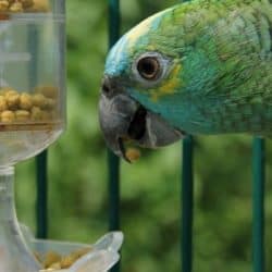
If you haven’t learned already millet is the Mars candy bar of bird treats which is why birds love it. We recommend it as a high-value training treat because of its fatty content but I decided during what we hope is Popcorn’s recuperation process to give her all of the millet she wants. I am giving her half a millet spray about six or 8 inches long on a piece of newspaper on the floor and let her have at it until she’s bored and/or full. I talked about this with Dr. Byron and he agreed with me. First of all because we really don’t know what her life expectancy will be – let her have the Mars bar.
But if you drill down on the idea, millet is packed with calories which makes it fattening. Illness, just like molting, reproduction, and flight, is a serious draw on calories. We benchmark her weight now every Friday when we visit Dr. Byron before and after the tap (today Friday, March 4, we pulled 7 mL total out of both sides of the abdomen which is one less mL than we pulled last Friday). We’re hoping this is a trend and will know next Friday. The millet will clearly add to her weight like I said but at this point we don’t care because we want it to be part of the overall mix of her treatment
The following is a comment to last week’s chat about human lungs and my ensuing answer (rant):
Thanks, Mitch, a very good topic that I thought I was up on… but ‘the German study’ was the last I’d heard. Many years ago I had a party and burned tons of candles all night. Once in bed, the carbon monoxide alarm went off and I called the fire company. When they were in, one turned to the parrots and said, Well you know the level isn’t too bad because they’re still standing.
Obviously, I don’t want to use my guys as a detector, and I’ve never lit many candles at once again. I also seek soy-based candles (about the only thing soy is good for, IMHO) vs. paraffin.
I’m so sorry to hear of Enki’s owner’s illness, and I hope she will look into the other home-based sources of irritants like those you suggested. Sending prayers for you.
Editorial (the rant):
Candles have no place in a home with birds – In 1842, Julius Robert Mayer discovered the “Law of Conservation of Energy”. In its most compact form, it is now called the First Law of Thermodynamics: energy is neither created nor destroyed. In lay terms – for a candle to produce heat and light energy it needs fuel which is the (paraffin) wax and the oxygen in the air. Every candle lit in the same room as a bird regardless if they are “bird-safe” is depleting oxygen from that room.
Cockatiel Sick with Psittacosis intake by Dr. Ross Perry specialist exotic avian vet
I have no scientific evidence but – much like dogs can sniff out human tumors and explosives the size of a grain of sand packaged tightly in luggage, I believe in my heart of hearts birds can feel the diminished oxygen from a single candle flame. Most candles usually do not lead to a bird’s death but are probably instrumental in reducing the quality of life for the animal while lit.
end (rant)
Which got me thinking about other ways your bird’s lungs could get damaged.
Aspergillosis
is the front runner of common fungal infections found in our pet birds. It’s got two personalities, the first usually seen in young birds who are been exposed to lots of spores of the Aspergillus fungus.
Mature birds are more susceptible to the Aspergillus fungus found in contaminated water, food, nesting material and/or poor ventilation. Vitamin A deficiency can enable the disease which additionally hits the bird’s lower respiratory tract. This is where the trachea, syrinx (voice organ), and bronchi may be affected along with the lungs and air sacs.
If your bird suffers from labored breathing or voice changes and it appears it has been affected by low-energy it may have Aspergillosis. The good news is this disease is treatable with antifungal drugs if caught in the early stages. The best defense against aspergillosis is plenty of fresh air ventilated in the cage or aviary area along with the right nutritional regime.
Bird flu
Although we associate Avian Influenza (Bird Flu) with the price of eggs and chicken, our pet birds are susceptible as well. This disease has caused the Centers for Disease Control and Prevention (CDC) to ban the importation of pet birds from certain countries in Africa, Europe, and Asia where avian influenza has been reported.
Birds transfer this disease by direct contact with respiratory secretions and feces from an infected bird. Birds can be infected but you may never know it. Other birds will just fall over dead. Infected birds may appear to have a compromised respiratory system, loss of appetite and swelling of the head, nasty goo discharge from the eyes, and really runny poop. If you think your bird has the bird flu it should be quarantined ASAP.
Macaw Asthma
It never ceases to amaze me that when people talk about macaws they are oblivious to the fact that there are 17 up to maybe 30 macaws if you take into account hybrids with sizes ranging from nine or 10 inches (Noble macaw) to almost 4 feet (Hyacinth macaw). Blue and gold macaws are especially susceptible to Macaw Respiratory Hypersensitivity (Macaw Asthma).
This makes the case against maintaining a flock with both South American and Australian and/or African birds. Cockatoos and African Greys that produce a powder-down can possibly trigger this disease. Much like Enki and last week’s story, a macaw would benefit from improved air quality through HEPA air filters. The biggest problem with this disease is the scarring of the bird’s lungs reducing their propensity for an activity like flying which reduces their overall health.
Psittacosis
How can you tell if the bird has Psittacosis? It’s important to note that – Chlamydia psittaci can affect not just pet caged birds but wild birds, chickens, ducks, pigeons and turkeys. It is also a Zootonic disease which means that it can be transferred to human beings making it even more insidious. Once infected the disease may not present itself for a few days or a few weeks.
If your bird looks tired, shivers, is losing weight (there’s that weighing regularly thing again) labored breathing or diarrhea this could be the cause. Infection can actually be latent which means they appear healthy but not show signs of infection into the future. The bird harbors that Chlamydia psittaci bacteria can in fact get rid of the organism intermittently even continuously for months. If your bird is not being fed properly, in an overcrowded cage environment, is laying eggs, it may shed the bacteria through its poop and its runny nose. The bacteria can actually remain infective for months.
Newcastle Disease
Ah – Newcastle Disease – triggered by a virus and although poultry farmers are concerned about it, caged bird keepers having pet birds would be well advised to be on the lookout for this disease.
It can manifest itself in many systems from head bobbing to wing drooping to bright yellow green diarrhea and body spasms leading to paralysis and sudden death. No care or treatment and your bird dies. We advocate that you report the death to the authorities thereby paying your due diligence.
Respiratory Parasites
And lest we forget Respiratory Parasites – a.k.a. air sac mites found throughout the entire respiratory system of an infected bird.
We see this a lot in canaries and Gouldian finches
Birds being birds disguising diseases, you may never know your bird suffers from this malady if it has a mild infection and having infection may cause your bird to exhibit labored breathing with rustling noises and clicking when sneezing and tail bobbing.
If you start picking up your bird and playing with it making it exercise you’re only going to make things worse. If the infection is pervasive your bird may die. If you’re lucky your veterinarian will prescribe an anti-parasitic that you need to give your bird on a regular basis.
another outstanding look at how birds lungs work
Gapeworms
Just the sound of “Gapeworms” sounds awful. They are parasites that will reside in the wall of your bird’s trachea favoring finches and canaries. The disease gets its name from birds that are “gaping for air”.
Sarcocystosis
And last but certainly not least Sarcocystosis is caused by a protozoan parasite that invades the body’s soft tissues and forms cysts in the respiratory tract, kidneys, nervous tissues, and eventually in the muscles. It affects cage birds primarily kept in the southern portion of the United States. Contaminated food is a trigger, foods that may be infected from insect feces like cockroaches or by rats feces showing up in the bird food.
It is treatable and preventable by keeping your bird food in a sealed container.
the crocodile thing
We all assume Mother nature designed avian lungs for the concept of flight but based on studies published as recently as 2010 and 2013 biologists determined crocodiles and alligators have similar respiratory systems to birds.
The human respiratory system probably occupies about 5% of the human body versus the avian respiratory system representing 20% of the birds volume.
Feathered factoids:
You and I take 12 to 16 breaths a minute – a greenwing macaw takes 30 to 40 breaths a minute – a canary 60 – 200 breaths a minute – The design of avian lungs allow them to sing and fly simultaneously –
Birds are actually descendants of dinosaurs and crocodiles are descendants of dinosaurs (the closest living relatives) crocodiles and related species have a similar respiratory system to birds.- one way airflow.
short answer
Figure 13: Diagrammatic representations of the crocodilian (A) and avian (B) lungs in left lateral view with colors identifying proposed homologous characters within the bronchial tree and air sac system of both groups. The image of the bird is modified from Duncker (1971). Abbreviations: AAS, abdominal air sac; CAS, cervical air sac; CRTS, cranial thoracic air sac; CSS, caudal sac-like structure; CTS, caudal thoracic air sac; d, dorsobronchi; GL, gas-exchanging lung; HS, horizontal septum; IAS, interclavicular air sac; L, laterobronchi; NGL, non-gas-exchanging lung; ObS, oblique septum; P, parabronchi; Pb, primary bronchus; Tr, trachea; v, ventrobronchi.
ref: Schachner ER, Hutchinson JR, Farmer C. (2013) Pulmonary anatomy in the Nile crocodile and the evolution of unidirectional airflow in Archosauria. PeerJ 1:e60 https://doi.org/10.7717/peerj.60
long answer
The information below although rather geeky does a good job of explaining the relationship between birds, crocodiles and alligators lungs
.

Figure 2. Archosaurs evolution tree – http://mappingignorance.org/author/mireia-altimira/
Alligator respiratory system Crocodilian lungs are distinct from bird lungs and are thought to have an alveolar-arterial blood gas exchange. However, the topography of the intrapulmonary bronchus and of the first bronchi is similar in birds and crocodilians.
-
The cervical ventral bronchus (CVB), pictured in green in Figure 4, is similar to the birds’ abdominal bronchus.
-
The three dorsobronchi (blue in Figure 4) spiral toward the apex of the lung like in birds. Farmer and Sanders have also discovered that the dorsal bronchi connect to each other and to the CVB through numerous parabronchi with diameters between 1 and 1.5 mm.
-
The small orifice to caudoventral bronchi (arrows, Fig. 1C) is placed opposite the orifice of the dorsal bronchi.
These anatomical similarities lead to the hypothesis that airflow in alligators might also be unidirectional. However, previous studies did not identify unidirectional flow in alligators’ lungs and this kind of flow was linked to the existence of abdominal air sacs and incompatible with the hepatic piston mechanism of ventilation of crocodilians.
Experiments
Three different sets of experiments were carried out to prove that airflow in alligator lungs is unidirectional. First, two flow-meters were implanted in the CVB (green in Figure 4) and in one of the dorsal bronchus (blue in Figure 4) of four alligators. Their lungs were then artificially ventilated, observing that air in the CVB moved in a cranial-to-caudal (head-to-tail) direction, while air in the dorsal bronchus moved in a ventrolateral (ventral part away from the midline) to dorsomedial (toward the back and near the midline) direction during both expiration and inspiration (Figure 4, F and G).
Next, the airflow in the CVB during normal breathing was monitored in six alligators. And finally, the flow was also visualized by filling an excised lung with a saline solution that contained fluorescent microspheres. This last test also allowed the characterization of the flow in the parabronchi, confirming that it is unidirectional.

Airflow in alligator lungs. Computed tomography images (left) show the hairpin turn (blue arrow) into the CVB (v) in the coronal (A) and medial sagittal (B) views. The lateral sagittal view shows the larger ostia to the dorsal bronchi (C), some of the ostia of ventral and lateral bronchi (blue arrows), a parabronchus (blue arrowheads), and the dorsal bronchus in which flow was recorded (d). The axial view (D) shows the bifurcation of the primary bronchi. An oblique dorsal view of the major bronchi is shown in (E). A simplified view shows airflow during inspiration (F) and exhalation (G) in the trachea, CVB, dorsal bronchus, and parabronchi. Hazelhoff’s model of exhalation (H) and inspiration (I). ipb, intrapulmonary bronchus; le, guiding dam; ve, vestibulum; v, ventrobronchus; m, mesobronchus; p, parabronchus; d, dorsobronchus; x,y, sites of constriction. | Credit: Sanders et al (2010)
In short, all three methods confirm that airflow through the lungs of alligators is unidirectional as in birds, since the flow during the transition from inspiration to expiration continued rather than dropping to zero, as would be the case of a tidal flow.
written by mitch rezman approved by catherine tobsing approved by nora caterino
References
- C. G. Sanders, K. Farmer, Unidirectional Airflow in the Lungs of Alligators, Science 327, 338 (2010)
Author Profile
Latest entries
 Feeding Exotic BirdsDecember 29, 2025How to Switch or Convert Your Bird From Seeds to Pellets: Real-Life Case Studies and Practical Guidance
 Feeding Exotic BirdsDecember 16, 2025A Practical, Budget-Smart Guide to Feeding Birds Well
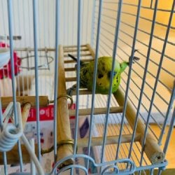 Bird EnviornmentsDecember 7, 2025Understanding Budgie Cage Bar Orientation: Myths, Realities & Practical Solutions for Vertical-Bar Bird Cages
 Feeding Exotic BirdsDecember 5, 2025How Dr. T.J. Lafeber Rewrote the Future of Pet Bird Nutrition