Last Updated on by Mitch Rezman
INTRODUCTION
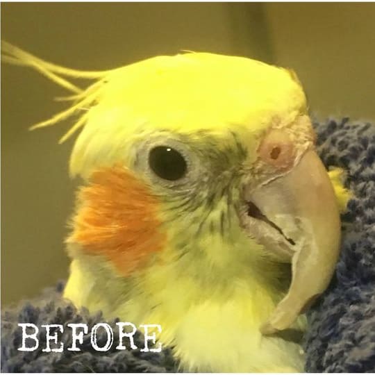
Rhamphorthotics, or correction of developmental abnormalities of the beak of juvenile psittacines has been well described by Clipsham and others. 1 2,3 Techniques are described for correction of “Scissors beak” or lateral deviation of the premaxilla or maxillary beak and for bradygnathism of the maxillary beak.
A simplified and more practical technique will be described here utilizing many of the same materials. A more invasive technique has also been described utilizing a pin placed through the frontal bone and rubber bands to pull the beak into alignment. 4
Developmental abnormalities of the beak are frequently seen in hand fed psittacines. They may result from improper handling or feeding techniques and malnutrition may increase the incidence. 5,6 Their occurrence however often cannot always be explained.
Laterial Deviations of the Maxillary Beak
Lateral deviations of the maxillary beak may on occasion be present at the time of hatching but more frequently become apparent at 2-4 weeks of age. If detected early they can usually be corrected by physical therapy in which finger pressure is used to push the beak back into a normal position. Self correction may occur.
The deviation may be to the right or the left. In the majority of cases the deviation affects only the keratin of the maxillary tip. In some cases the entire maxillary beak (rostrum maxillaris) is deviated. Rarely the frontal hinge and possibly the frontal bone may be affected. These deviations will result in uneven wear and overgrowth of the beak due to poor opposition of the occlusal surface of the mandibular beak and the serrated surface of the occlusal surface of the maxillary beak.
Very young chicks should be monitored closely for signs of development of lateral deviations. A number of factors may be contributory. The occlusal surface of the mandible may be uneven. A frenulum like thickening of keratin is often present in chicks and is often off center. This area of thicker keratin will wear more slowly and cause a high point which can contribute to pushing the soft beak to one side.
Handling or beak cleaning techniques may also be contributory. Care takers often wipe the beak from the lateral commisures to the tip putting slight but repeated pressure on the soft beak. In feeding if the caretaker holds the head with finger pressure on the lateral commisures of the beak this can also be contributory to compression of the tip of the beak. This compression makes the occlusal surface more pointed and more narrow. As the beak grows and becomes calcified, the malformation becomes self perpetuating as the uneven wear of occlusal surfaces contributes to continued or even potentiated lateral deviation.
Correction of beak malformations by physical therapy
Physical manipulation should always be the first option and can be successful up until the time that the beak becomes so calcified that it is not movable by finger pressure. Prior to beak therapy the beak must be trimmed with a grinding wheel (Dremel, Racine, WI) so that the occlusal surfaces of both the maxillary and mandibular beaks are normal. Repeated trimming may be necessary. In very small chicks the occlusal surface of the mandible is trimmed with cuticle nippers.
For physical therapy in very young chicks, the beak is manipulated with the thumb and fore finger bending the tip of the maxilla toward the center and over correcting slightly. To be effective the therapy will be somewhat painful to the chicks. Soothing the chick following therapy will prevent fear of approach. If possible the chick should also be fed after therapy. Therapy can be once daily or more often if needed.
Mild cases may be resolved by grinding. A conical, wood grinding tip is used. The occlusal surface of both the maxillary and mandibular beak is ground. If the beak can be closed with the tip of the grinding wheel between the surfaces can both be ground into a shape which will allow the beak to close properly after trimming. If bleeding occurs grinding should be discontinued. The beak can be ground weekly as needed to correct mild cases.
As in cases of lateral deviation, this defect can be corrected by physical therapy in very young chicks. There is a soft, cartilaginous flange present on the lateral occlusal surfaces of the maxillary beak in hatchling cockatoos. As they become older, this flange may pull the tip of the maxilla inward. This flange can be trimmed with cuticle scissors and should be trimmed in any abnormal chick. Perhaps it would be worn off by the parents in chicks in the nest.
After trimming the flange, pull the tip of the maxilla over the front of the mandible and hold for a short time in place. If it will not easily extend over the mandible the tip should be gently pulled forward taking care not to crease the cranial aspect of the maxilla. In cockatoo chicks the mandible is also often compressed laterally causing it to become pointed and elongated. This contributes to the early disparity in length of the beaks and may aggravate mild cases of bradygnathism. If compressed the mandible should be ground with a grinding wheel and the cranial aspect widened by pressing the thumb inside the mandible and spreading it laterally.
Correction of beak deformities by acrylic device
Simplified and practical techniques of correction of beak malformations provide correction at reduced cost. For best results the repair should be initiated shortly after calcification of the beak makes it non-pliable. If corrected prior to placement of the bird into a cage, the rate of failure is very low and the time required for correction is short.
The bird should be fasted sufficiently to empty the crop of formula, or the procedure should be done prior to the morning feeding. Alternatively the bird can be given a small feeding early in the morning prior to surgery. The bird should be anesthetized with isoflourane or other suitable anesthetic. Application of the device is impossible in an unanesthetized bird as it will bite the acrylic prior to hardening. Endotracheal or abdominal air sac intubation is necessary.
Clipsham described a successful technique but it is difficult, time consuming and requires many steps for completion. It is possible that this technique or a modification thereof may e needed in very severe cases. The Visible light curing acrylic is designed for dental use and produces no heat upon curing. (Sunschein Restorative, Henry Schein Inc, Port Washington, NY 11050) The acrylic which is hardened by application of an intense source of ultraviolet light. The acrylic hardens within 10 seconds to a minute depending on the thickness of the acrylic.
For a more durable application the acrylic can be applied in small sections or layers and hardened between applications. Eye protection in the form of a shield or glasses must be utilized to prevent retinal damage. Handling acrylic with gloved or moistened hands helps to prevent sticking of acrylic to the hands. Cooling the acrylic may reduce handling difficulties.
Clipsham’s technique of preparation of the beak for application of acrylic includes scoring of the keratin and roughing of the surface.
In this simplified technique, a stainless steel intramedullary pin or an appropriately sized hypodermic needle can be placed through the beak to anchor the acrylic. The pin is driven through the beak from the cranial surface to the lateral wall on the side to which the maxilla deviates. The pin is placed approximately 1-3 mm below the occlusal surface. An acrylic ramp is then constructed to redirect the maxilla through an inclined plane.
It is not necessary to extend this ramp very high as the force of the maxilla sliding toward center can be achieved with a gently sloping incline. The acrylic ramp must however be extended far enough laterally to the side which the beak deviates so that the tip of the maxilla cannot be placed lateral to the acrylic as this will aggravate the problem. The steeper the ramp, the move rapidly the beak will correct. Over correction may occur. If the device cannot be monitored closely, or as a routine measure to prevent over correction another usually smaller ramp can be placed on the contralateral side. The lowest point between the two ramps should be off center in the direction needed for correction.
After construction of the ramp it can be smoothed and the shape and slope refined with the grinding wheel.
The technique described here is simple and rapid requiring minimal materials and time It usually results in correction in 1-2 weeks. Some may require longer and rarely a device may be lost and require replacement. Rate of loss of the device is low in young macaws. Putting birds into cages with the device in place should be avoided to prevent fracture of the device when biting the wire or bars.
Anesthesia is not required for removal. Grind a line along the occlusal surface and ventrally along the midline through the acrylic. The device can then be easily removed by hand and the pin removed. Covering or sealing the hole is not necessary. After removal, smooth the occlusal surface of the mandibular beak with the grinding wheel. Occasionally the beak may fracture to the hole. As the hole is close to the occlusal surface it can often be ground down to a smooth surface initially or within a few weeks.
A similar technique is used for correction of mandibular prognathism. Place the bird under isoflourane anesthesia administered by endotracheal intubation or air-sac canulation. Grind the beak to as close to normal as possible. The artificial extension of the maxillary beak over the mandibular beak directs the growth of the chick in a proper direction and also prevents contraction of the ligaments of the nasofrontal hinge.
Grind the beak into normal shape with a grinding wheel. In Clipsham’s technique, one or more grooves are cut or ground in the keratin of the maxillary beak and then roughens the external keratin. In this technique a stainless steel intramedullary pin or appropriately sized hypodermic needle is placed through the tip of the maxillary beak from one side to the other which serves to attach the acrylic. The acrylic is then applied in layers extending over approximately half of the three outer surfaces of the beak, incorporating the pin.
Alternatively the acrylics can be rolled into a cone. Push the acrylic up onto all three external surfaces of the.The acrylic tip is extended down the tip of the beak and farther downward approximately 1/4 to 1/2 inches depending on the size of the bird. A long extension on the device is not necessary if the acrylic is formed so that the tip is curved cranially and directed far enough so that is impossible for the bird to put it inside the mandible.
The tip can be widened adding stability and making it more difficult to place inside. The shape of the occlusal surface should match that of the mandible to prevent lateral deviation. In chicks which also have a lateral deviation or start to develop one secondarily, a device like the one used for correction of lateral deviations can be applied to the center of the mandible to prevent deviation to one side or the other. The acrylic may be light cured in sections or as a unit. Extend the cone rostrally at approximately a 60 to 90 degree angle?
The appliance must be of uniform thickness with sufficient thickness to prevent fracture but not to add excess weight. The surfaces should be smooth and the edges confluent with the beak to prevent food retention. Smooth rough spots with a grinding wheel. It should be impossible or difficult to place the appliance tip into the mandibular beak while the bird is under anesthesia.
Conclusion
These simple techniques for correction of developmental abnormalities should make correction rapid and economical allowing resolution of the problem early enough to increase the chance of success and avoid interference with weaning.
References:
1. Altman, Robert B., Beak Repair, Acrylics, In; Avian Medicine and Surgery, Altman, et.al., W.B. Saunders Co., Philadelphia, PA, 1996 – pp. 787-799.
2. Clipsham, Robert, Surgical Beak restoration and correction, Proc. Assoc Avian Vet, Seattle, 1989, pp.164-176.
3. Clipsham, Robert, Beak Repair, Rhamphorthotics in; Avian Medicine and Surgery, Altman, et.al., W.B. Saunders, Philadelphia, PA, 1996 – pp 773- 786.
4. Martin, Howard and Branson W. Ritchie, Orthopedic Surgical Techniques, In Avian Medicine: Principles and Application, Ritchie, Branson W., Greg J. Harrison and Linda R. Harrison, Wingers Publishing, Inc., Lake Worth, FL., pp 1137 – 1169.
5. Speer BL., Pediatric medical management, in The Large Macaws, Their Care, Breeding and Conservation Abramson J, Speer BL, Thomson JB (eds); Raintree Publications, Fort Brigg, CA, 1995, pp 361-372.
6. Wolf S., Clubb S.L., : Clinical management of beak malformations in handfed psittacine chicks, in Schubot, Clubb K, Clubb SL (eds) Psittacine Aviculture, Perspectives, Techniques and Research, Avicultural Breeding and Research Center, Loxahatchee, FL, 1992 pp- 17/1-17/11.
Courtesy of Pretty Bird
Author Profile
Latest entries
 PigeonsJune 20, 2025How Do Parrots Thrive in Cities Outside Their Native Habitats?
 Feeding Exotic BirdsJune 20, 2025Is Corn On the Cob Safe for Pet Birds?
 Bird & Parrot AnatomyJune 19, 2025Would You Like a Comprehensive Guide for Training Your Bird?
 Feeding Exotic BirdsJune 19, 2025Here Are Three (Video) Strategies to Help Your Bird Forage
